Thoracic aorta, 1825 artwork

![]()
Wall Art and Photo Gifts from Science Photo Library
Thoracic aorta, 1825 artwork
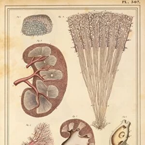
Thoracic aorta. Dissection of the chest cavity with the heart and lungs and other organs removed, showing the thoracic portion of the aorta (red), the main body artery. This artery carries oxygenated blood from the heart to the rest of the body. The arteries branching off it are shown here. This anatomical artwork is plate 226 from volume 4 of Manuel d anatomie descriptive du corps humain (1825). This 5-volume anatomy atlas was produced by French physician and surgeon Jules Germain Cloquet (1790-1883). The illustrations were by Haincelin. Volume 4 illustrated the anatomy of the circulatory and respiratory systems
Science Photo Library features Science and Medical images including photos and illustrations
Media ID 9273415
© SCIENCE PHOTO LIBRARY
1825 Anatomical Artwork Anatomical Illustration Anatomy Atlas Anterior Aorta Arterial System Arteries Blood Vessels Branches Chest Chest Cavity Dissected Dissection French Frontal Haincelin Jules Germain Cloquet Oxygenated Blood Ribs Thoracic Thorax Vascular Volume 4 Volume Iv Artery Blood Vessel Circulatory System Ribcage
EDITORS COMMENTS
This artwork, titled "Thoracic aorta" takes us back to the 19th century when French physician and surgeon Jules Germain Cloquet produced his remarkable anatomy atlas. Plate 226 from volume 4 of Manuel d'anatomie descriptive du corps humain (1825), this print showcases the intricate network of blood vessels within the thoracic portion of the aorta. The dissection reveals a mesmerizing sight: the heart and lungs have been removed, allowing us to focus solely on this vital artery that carries oxygenated blood from the heart to every corner of our bodies. The vibrant red color emphasizes its significance as it branches off into smaller arteries, ensuring proper circulation throughout our system. With meticulous detail, Haincelin's illustrations showcase not only the anterior view but also provide an insight into how these blood vessels intertwine with ribs and other structures within the chest cavity. This historical piece serves as a testament to both medical advancements and artistic brilliance in capturing anatomical accuracy. As we gaze upon this extraordinary artwork, we are reminded of how far we have come in understanding our own bodies. It is through such visual representations that we can appreciate the complexity and beauty hidden beneath our skin.
MADE IN THE USA
Safe Shipping with 30 Day Money Back Guarantee
FREE PERSONALISATION*
We are proud to offer a range of customisation features including Personalised Captions, Color Filters and Picture Zoom Tools
SECURE PAYMENTS
We happily accept a wide range of payment options so you can pay for the things you need in the way that is most convenient for you
* Options may vary by product and licensing agreement. Zoomed Pictures can be adjusted in the Cart.