Kidney anatomy, 1825 artwork

![]()
Wall Art and Photo Gifts from Science Photo Library
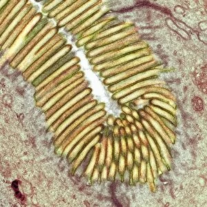
Kidney anatomy, 1825 artwork
Kidney anatomy. Artworks of: superficial renal veins (blue, upper left); renal tubules (top right); arteries (red) in sectioned kidney (centre left); renal vessels (wax injection, lower left); ureter (white) in sectioned kidney (bottom centre); kidney stones in ureter (lower right). This anatomical artwork is plate 307 from volume 5 of Manuel d anatomie descriptive du corps humain (1825). This 5-volume anatomy atlas was produced by French physician and surgeon Jules Germain Cloquet (1790-1883). The illustrations were by Haincelin. Volume 5 illustrated the anatomy of the digestive, secretory and reproductive organs
Science Photo Library features Science and Medical images including photos and illustrations
Media ID 9222853
© SCIENCE PHOTO LIBRARY
1825 Abdomen Anatomical Artwork Anatomical Illustration Anatomy Atlas Arteries Cortex Cortical Dissected Dissection Excretory System French Haincelin Histological Histology Jules Germain Cloquet Kidney Kidney Stone Medulla Organs Renal Stones Superficial Urinary Urinary System Veins Volume 5 Volume V Section Sectioned
MADE IN THE USA
Safe Shipping with 30 Day Money Back Guarantee
FREE PERSONALISATION*
We are proud to offer a range of customisation features including Personalised Captions, Color Filters and Picture Zoom Tools
SECURE PAYMENTS
We happily accept a wide range of payment options so you can pay for the things you need in the way that is most convenient for you
* Options may vary by product and licensing agreement. Zoomed Pictures can be adjusted in the Cart.