Small intestine anatomy, artwork

![]()
Wall Art and Photo Gifts from Science Photo Library
Small intestine anatomy, artwork
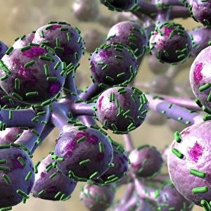
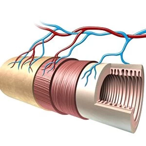
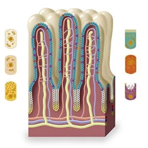
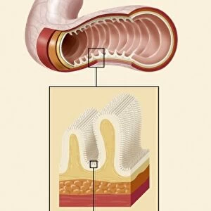
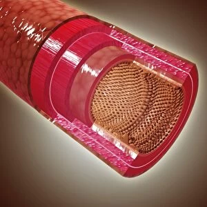
Small intestine. Artwork of a section through the duodenum, part of the small intestine. The small intestine is where digestion begins and nutrients are absorbed into the blood. The lumen (white, centre) is lined with villi, the folds in the intestinal surface that greatly increase the surface area for absorption. The smooth muscle layer beneath the villi (orange) is used to mix the food as it passes through the lumen. Surrounding this is circular (white lines) and longitudinal (white dots) muscle tissue, the peristaltic action of which pushes food along the intestine
Science Photo Library features Science and Medical images including photos and illustrations
Media ID 6345231
© FRANCIS LEROY, BIOCOSMOS/SCIENCE PHOTO LIBRARY
Alimentary Canal Bowel Digestive System Duodenal Duodenum Gastrointestinal Tract Intestinal Intestines Lumen Small Intestine Villi
EDITORS COMMENTS
This artwork showcases the intricate anatomy of the small intestine, specifically a section through the duodenum. The small intestine is an essential organ where digestion commences and vital nutrients are absorbed into our bloodstream. In this print, we can observe the lumen at the center, depicted in white, which is lined with numerous villi. These tiny folds on the intestinal surface play a crucial role in maximizing absorption by significantly increasing its surface area. The orange layer beneath these villi represents smooth muscle tissue responsible for mixing food as it traverses through the lumen. Surrounding this layer, we can see circular white lines and longitudinal white dots representing muscle tissues that facilitate peristaltic action - pushing food along our intestines. Set against a striking black background, this illustration beautifully captures both artistry and biology. It offers us a glimpse into one of nature's wonders: our own digestive system. This print not only serves as an educational tool but also highlights the marvels of human anatomy. With its attention to detail and accuracy, Science Photo Library has once again delivered an exceptional piece that appeals to those fascinated by science and health alike. Whether you're studying biology or simply intrigued by how your body works, this print provides insight into our complex gastrointestinal tract while showcasing its beauty in artistic form.
MADE IN THE USA
Safe Shipping with 30 Day Money Back Guarantee
FREE PERSONALISATION*
We are proud to offer a range of customisation features including Personalised Captions, Color Filters and Picture Zoom Tools
SECURE PAYMENTS
We happily accept a wide range of payment options so you can pay for the things you need in the way that is most convenient for you
* Options may vary by product and licensing agreement. Zoomed Pictures can be adjusted in the Cart.