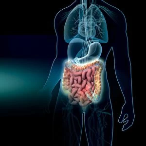
Intestinal anatomy, artwork

![]()
Wall Art and Photo Gifts from Science Photo Library
Intestinal anatomy, artwork
Intestinal anatomy. Computer artwork showing the layers of the small intestine. The central space (lumen) is surrounded by the mucosa (beige, folded), which has numerous folds (villi). Surrounding this is the submucosa (beige) and then a layer of circular muscle (brown), followed by a layer of longitudinal muscle. The outer layer (beige, left) is the serous membrane (serosa). The arteries, which supply blood to the intestine, and veins that carry nutrients, absorbed during digestion, away from the intestine, can also be seen
Science Photo Library features Science and Medical images including photos and illustrations
Media ID 6324503
© HENNING DALHOFF / SCIENCE PHOTO LIBRARY
Arteries Fold Folds Gastroenterology Gastrointestinal Intestinal Intestine Layer Layers Longitudinal Muscle Lumen Mucosa Muscles Physiological Physiology Small Intestine Structures Sub Mucosa System Vascular System Veins Villi Villus Artery Blood Supply Serosa Vein
EDITORS COMMENTS
This artwork showcases the intricate layers and structures of the small intestine, providing a glimpse into the fascinating world of human anatomy. The computer-generated illustration highlights the complexity and functionality of this vital organ within our digestive system. At its core, we see the central space known as the lumen, encased by the mucosa layer with its distinctive folds called villi. These folds play a crucial role in increasing surface area for nutrient absorption during digestion. Surrounding the mucosa is the submucosa layer followed by a layer of circular muscle, responsible for propelling food through peristaltic contractions. The outermost layer, known as serous membrane or serosa, provides protection to these underlying layers. The image also reveals an intricate network of arteries and veins that supply blood to and from the intestine respectively. This vascular system ensures efficient delivery of nutrients absorbed during digestion throughout our body. With its detailed portrayal of anatomical structures such as arteries, veins, muscles, and folds, this print serves as both an educational tool for students studying biology or gastroenterology and a visually stunning piece of art that celebrates our complex physiology.
MADE IN THE USA
Safe Shipping with 30 Day Money Back Guarantee
FREE PERSONALISATION*
We are proud to offer a range of customisation features including Personalised Captions, Color Filters and Picture Zoom Tools
SECURE PAYMENTS
We happily accept a wide range of payment options so you can pay for the things you need in the way that is most convenient for you
* Options may vary by product and licensing agreement. Zoomed Pictures can be adjusted in the Cart.