Dividing cancer cell, SEM C014 / 0361

![]()
Wall Art and Photo Gifts from Science Photo Library
Dividing cancer cell, SEM C014 / 0361
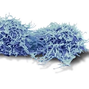
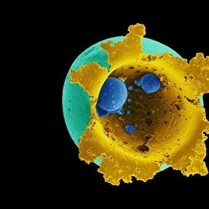
Dividing cancer cell. Coloured scanning electron micrograph (SEM) of a colorectal cancer cell undergoing mitosis (nuclear division) and splitting into two daughter cells (left and right). Here, it is in late telophase, the final stage before cell division (cytokinesis) and the two daughter cells are still connect by a cytoplasmic bridge (horizontal, centre). Bacteria (rod-shaped) can also be seen on the cells. Magnification: x2000 when printed at 10 centimetres wide
Science Photo Library features Science and Medical images including photos and illustrations
Media ID 9219171
© STEVE GSCHMEISSNER/SCIENCE PHOTO LIBRARY
Bacteria Bacterium Bowel Cancer Cancer Cell Cancerous Cell Cycle Colon Cancer Colonic Colorectal Cancer Colored Culture Cytological Cytology Cytoplasmic Bridge Daughter Cells Dividing Division Gastroenterological Gastroenterology Gastrointestinal System Intestinal Intestines Large Intestine Malignancy Malignant Microvilli Microvillus Mitosis Mitotic Oncological Oncology Replicating Replication Telophase Tract Villi Villus Abnormal Cells Condition Disorder Unhealthy
EDITORS COMMENTS
This photo print, captured by the scanning electron microscope, showcases a dividing cancer cell in all its intricate detail. The image depicts a colorectal cancer cell undergoing mitosis, the process of nuclear division, as it splits into two daughter cells. In this late telophase stage, just before cell division (cytokinesis), the two daughter cells remain connected by a cytoplasmic bridge at the center. The vibrant colors bring attention to not only the malignant nature of this abnormal duo but also highlight the presence of rod-shaped bacteria on the surface of these cells. With a magnification of x2000 when printed at 10 centimeters wide, every minute feature is revealed with astonishing clarity. This image holds significant importance in both biology and medicine as it provides valuable insights into cellular replication within intestinal tissues affected by colorectal cancer. It sheds light on various aspects such as microvilli and villi structures present in our gastrointestinal system. While visually striking, it serves as a reminder of the challenges posed by diseases like cancer and underscores ongoing efforts in oncology research to understand and combat these conditions effectively. This photograph captures not only scientific curiosity but also represents hope for advancements in healthcare that can lead to improved diagnosis and treatment options for patients worldwide.
MADE IN THE USA
Safe Shipping with 30 Day Money Back Guarantee
FREE PERSONALISATION*
We are proud to offer a range of customisation features including Personalised Captions, Color Filters and Picture Zoom Tools
SECURE PAYMENTS
We happily accept a wide range of payment options so you can pay for the things you need in the way that is most convenient for you
* Options may vary by product and licensing agreement. Zoomed Pictures can be adjusted in the Cart.