Home > Animals > Mammals > Muridae > Water Mouse
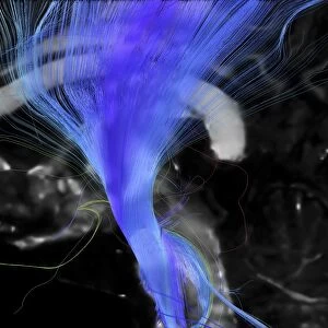
Brains white matter, DTI MRI scan

![]()
Wall Art and Photo Gifts from Science Photo Library
Brains white matter, DTI MRI scan
Brains white matter. Close-up of an area of the brain imaged using tract density imaging and 3D diffusion tensor imaging (DTI), a magnetic resonance imaging (MRI) technique. This frontal view of the centrum semiovale (white matter area) shows crossing areas between the corpus callosum (red), the corticospinal tract (blue) and the superior longitudinal fasciculus (green). The fibres are locally coloured red-green-blue if they are orientated in x-y-z alignment (left-right, posterior-anterior, inferior-superior). Diffusion tensor imaging (tractography) measures the direction of water diffusion, which in the brain reveals the orientation of nerve fibres
Science Photo Library features Science and Medical images including photos and illustrations
Media ID 9339875
© SHERBROOKE CONNECTIVITY IMAGING LAB/SCIENCE PHOTO LIBRARY
Brain Imaging Brain Scan Central Nervous System Cerebral Cerebrum Corpus Callosum Diffusion Tensor Imaging Dti Scan Fasciculus Fiber Fibers Fibre Fibres Imaging Technique Magnetic Resonance Imaging Mri Scan Mri Scanner Nerve Nerve Fibre Nerves Neural Pathway Neural Tract Paths Pathway Pathways Structural Tractogram Tractography White Matter Brain Neurological Neurology
FEATURES IN THESE COLLECTIONS
> Animals
> Mammals
> Muridae
> Water Mouse
EDITORS COMMENTS
This print showcases the intricate beauty and complexity of the human brain's white matter. Using advanced imaging techniques such as tract density imaging (TDI) and 3D diffusion tensor imaging (DTI), this frontal view of the centrum semiovale, a region in the brain's white matter, reveals a mesmerizing network of neural pathways. The image highlights crossing areas between three major tracts: the corpus callosum depicted in red, responsible for connecting both hemispheres of the brain; the corticospinal tract shown in blue, which plays a crucial role in motor function; and finally, the superior longitudinal fasciculus represented by green fibers that are associated with language processing and attention. Each fiber within this neural tapestry is locally colored using an RGB scheme to indicate its orientation along different axes. This technique allows researchers to visualize how water diffusion aligns with nerve fibers' directionality, providing valuable insights into their structural organization. With its black background enhancing contrast and vibrant colors illuminating every pathway, this print captures not only the anatomical intricacies but also represents cutting-edge advancements in medical technology. It serves as a reminder of how far we have come in understanding our own biology while offering endless possibilities for further exploration into neurological research and clinical applications.
MADE IN THE USA
Safe Shipping with 30 Day Money Back Guarantee
FREE PERSONALISATION*
We are proud to offer a range of customisation features including Personalised Captions, Color Filters and Picture Zoom Tools
SECURE PAYMENTS
We happily accept a wide range of payment options so you can pay for the things you need in the way that is most convenient for you
* Options may vary by product and licensing agreement. Zoomed Pictures can be adjusted in the Cart.