Broken upper arm bone, X-ray

![]()
Wall Art and Photo Gifts from Science Photo Library
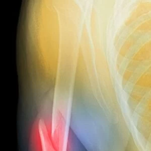
Broken upper arm bone, X-ray
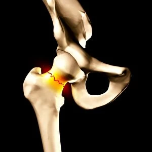
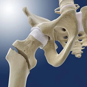
Broken upper arm bone. Coloured X-ray of a fracture (yellow) of the upper arm bone (humerus). The neck of the humerus has fractured, separating its head (upper right) from its shaft (below the head). This type of injury would require the bone to be pinned in place to heal. The humeral head connects (articulates) to the scapula (shoulder blade) to its left, forming the ball-and-socket shoulder joint. Above the scapula, lies the clavicle (collar bone) that articulates with the scapula. The ribs of the chest are at left
Science Photo Library features Science and Medical images including photos and illustrations
Media ID 6415508
© DU CANE MEDICAL IMAGING LTD/SCIENCE PHOTO LIBRARY
Ball And Socket Blade Bones Break Broken Clavicle Collar Diagnosis Diagnostic Displaced Displacement Fracture Fractured Front Frontal Humerus Injured Injury Joint Radiograph Radiography Radiology Scan Scapula Shaft Shoulder Upper Condition Disorder Health Care
EDITORS COMMENTS
This print from Science Photo Library showcases a vividly colored X-ray of a broken upper arm bone. The fracture, depicted in yellow, is located at the neck of the humerus, causing a separation between its head and shaft. Such an injury necessitates the placement of pins to facilitate healing. The significance of this image lies in highlighting the intricate anatomy and interconnectedness within our bodies. The humeral head connects with the scapula on its left side, forming the ball-and-socket shoulder joint that enables fluid movement. Above the scapula rests the clavicle, or collarbone, which articulates with it. Examining this X-ray also provides insight into medical diagnosis and treatment options for fractures. Radiography plays a crucial role in identifying bone disorders and assessing their severity accurately. In cases like these where displacement occurs, intervention becomes necessary to ensure proper healing. Moreover, this image serves as a reminder of our vulnerability to injuries and emphasizes the importance of healthcare professionals who specialize in radiology and orthopedics. By capturing such detailed visuals through diagnostic scans like frontal radiographs, medical experts can effectively diagnose conditions and develop appropriate treatment plans. In conclusion, Science Photo Library's striking print not only captures an injured upper arm but also sheds light on various aspects related to medicine, health care delivery systems, diagnostics processes involving radiology techniques while showcasing human anatomy intricacies beautifully captured by modern technology.
MADE IN THE USA
Safe Shipping with 30 Day Money Back Guarantee
FREE PERSONALISATION*
We are proud to offer a range of customisation features including Personalised Captions, Color Filters and Picture Zoom Tools
SECURE PAYMENTS
We happily accept a wide range of payment options so you can pay for the things you need in the way that is most convenient for you
* Options may vary by product and licensing agreement. Zoomed Pictures can be adjusted in the Cart.