Home > Animals > Insects > Mites > Follicle Mite
Follicle mite heads (SEM) C013 / 5120
 C013 / 5120")
![]()
Wall Art and Photo Gifts from Science Photo Library
Follicle mite heads (SEM) C013 / 5120
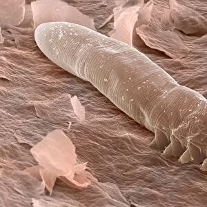
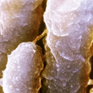
Follicle mite heads (Demodex folliculorum), coloured scanning electron micrograph (SEM) of follicle or eyelash mites protruding from a dissected human hair follicle. These mostly harmless parasites infest hair follicles (depressions in the skin which contain the roots of hairs) around the eyelids, nose and in the ear canals of humans. One follicle may contain up to 25 growing mites. They feed on oily secretions from the skins sebaceous glands, as well as dead skin cells. Infestation with follicle mites is usually symptomless, but allergic reactions to the mites in susceptible individuals can cause hair loss and acne. Magnification X530 at 10cm wide
Science Photo Library features Science and Medical images including photos and illustrations
Media ID 9195777
© POWER AND SYRED/SCIENCE PHOTO LIBRARY
Acne Allergen Allergy Arachnida Commensal Demodex Folliculorum Ectoparasite Eyelash Eyelash Mite Follicle Mite Hair Mite Mites Nymph Parasite Parasitic Skin
FEATURES IN THESE COLLECTIONS
> Animals
> Insects
> Mites
> Follicle Mite
> Animals
> Insects
> Mites
> Related Images
EDITORS COMMENTS
This print showcases the intricate world of follicle mite heads, specifically Demodex folliculorum. Through a colored scanning electron micrograph (SEM), we are granted a close-up view of these mostly harmless parasites protruding from a dissected human hair follicle. These minuscule creatures infest the hair follicles around our eyelids, nose, and ear canals. Within just one follicle, up to 25 growing mites may reside. Their diet consists of oily secretions from our sebaceous glands and dead skin cells. While infestation with these mites typically goes unnoticed, susceptible individuals may experience allergic reactions leading to hair loss and acne. The magnification level of this SEM image is an astonishing X530 at 10cm wide, allowing us to appreciate the intricate details of each individual mite head. This photograph serves as a reminder that even within the tiniest corners of our bodies exists an entire ecosystem teeming with life. Captured by POWER AND SYRED/SCIENCE PHOTO LIBRARY, this image not only highlights the scientific significance but also possesses an undeniable aesthetic appeal. It provides valuable insights into the world of parasitic arachnids while simultaneously evoking curiosity about our own bodies' complex interactions with nature's smallest inhabitants.
MADE IN THE USA
Safe Shipping with 30 Day Money Back Guarantee
FREE PERSONALISATION*
We are proud to offer a range of customisation features including Personalised Captions, Color Filters and Picture Zoom Tools
SECURE PAYMENTS
We happily accept a wide range of payment options so you can pay for the things you need in the way that is most convenient for you
* Options may vary by product and licensing agreement. Zoomed Pictures can be adjusted in the Cart.