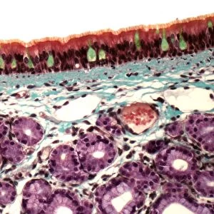
Typhoid nodules, light micrograph

![]()
Wall Art and Photo Gifts from Science Photo Library
Typhoid nodules, light micrograph
Typhoid nodules, coloured light micrograph. Section through a lymph node of a patient with typhoid, showing macrophages (large cells, pink) forming typhoid nodules. Typhoid is a disease caused by the bacterium Salmonella typhi and is transmitted by contaminated food and water. Macrophages normally engulf and destroy pathogens found in the lymph fluid. However, S. typhi is adapted to avoid destruction and infects the macrophage that engulfs it. The infected macrophages then aggregate within tissues to form typhoid nodules. If not treated with antibiotics, these can lead to internal bleeding and death
Science Photo Library features Science and Medical images including photos and illustrations
Media ID 6415234
© STEVE GSCHMEISSNER/SCIENCE PHOTO LIBRARY
Aggregation Bacteria Bacterial Bacterium Clumps Clusters Diagnosis Diagnostic False Colour Histopathological Histopathology Infected Infection Invasive Lesion Lesions Lymph Node Lymphocytes Macrophages Nodule Nodules Pathological Pathology Slice Tissue Abnormal Clustering Condition Disorder False Coloured Health Care Light Micrograph Light Microscope Section Sectioned Typhoid Unhealthy
EDITORS COMMENTS
This print showcases the intricate details of Typhoid nodules, providing a glimpse into the devastating effects of this bacterial disease. In this coloured light micrograph, we observe a section through a lymph node of a patient afflicted with typhoid. The prominent pink cells are macrophages, which typically engulf and eliminate pathogens in the lymph fluid. However, Salmonella typhi, the bacterium responsible for typhoid fever, has evolved to evade destruction and instead infects these macrophages. As depicted in this image, infected macrophages aggregate within tissues to form typhoid nodules. If left untreated with antibiotics, these nodules can lead to severe complications such as internal bleeding and even death. This visual representation sheds light on the invasive nature of S. typhi and its ability to cause significant tissue damage. Typhoid is primarily transmitted through contaminated food and water sources. Understanding its pathology is crucial for accurate diagnosis and effective treatment strategies. Through histopathological examination using techniques like sectioning and staining, medical professionals can identify characteristic clusters or clumps of infected macrophages within lymph nodes. Science Photo Library presents this remarkable image that merges science with artistry—a testament to their commitment in capturing visually stunning representations of various diseases and conditions affecting the human body.
MADE IN THE USA
Safe Shipping with 30 Day Money Back Guarantee
FREE PERSONALISATION*
We are proud to offer a range of customisation features including Personalised Captions, Color Filters and Picture Zoom Tools
SECURE PAYMENTS
We happily accept a wide range of payment options so you can pay for the things you need in the way that is most convenient for you
* Options may vary by product and licensing agreement. Zoomed Pictures can be adjusted in the Cart.