Skin layers, SEM

![]()
Wall Art and Photo Gifts from Science Photo Library
Skin layers, SEM
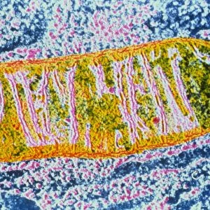
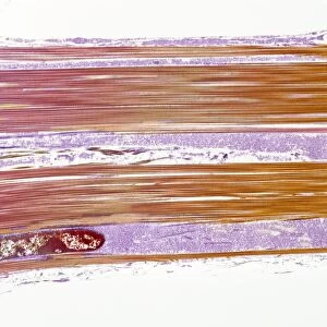
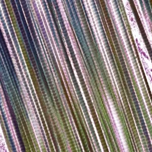
Skin. Coloured scanning electron micrograph (SEM) of a freeze-fractured section through human skin. At top is the stratum corneum (pale yellow) of the epidermis, a cornified layer composed of flattened, dead skin cells which form the surface of the skin. The dead cells from this layer are continuously being shed and replaced from cells from the living epidermal layer below (purple). The lowest layer seen here is the dermis (pale yellow and purple strands). This is a thick layer of fibrous connective tissue which supports and nourishes the epidermis. The skin is the bodys largest organ, accounting for around 15% of the bodys weight. Magnification unknown
Science Photo Library features Science and Medical images including photos and illustrations
Media ID 6455141
© STEVE GSCHMEISSNER/SCIENCE PHOTO LIBRARY
Cornified Layer Cross Section Dead Dermis Epidermis Freeze Fracture Freeze Fractured Layers Skin Stratum Corneum Surface Cells Section Sectioned
MADE IN THE USA
Safe Shipping with 30 Day Money Back Guarantee
FREE PERSONALISATION*
We are proud to offer a range of customisation features including Personalised Captions, Color Filters and Picture Zoom Tools
SECURE PAYMENTS
We happily accept a wide range of payment options so you can pay for the things you need in the way that is most convenient for you
* Options may vary by product and licensing agreement. Zoomed Pictures can be adjusted in the Cart.