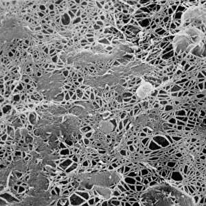
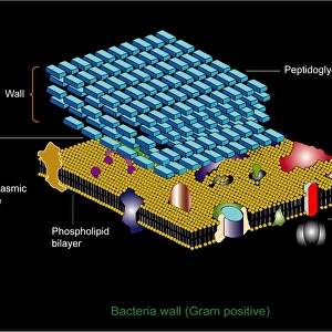
Protoplast showing cellulose microfibrils

![]()
Wall Art and Photo Gifts from Science Photo Library
Protoplast showing cellulose microfibrils
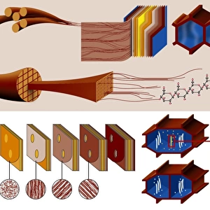
Scanning electron micrograph of the surface of a tobacco leaf protoplast, Nicotiana tabacum, showing cellulose microfibrils regrowing over the plasma membrane. A protoplast is a single intact cell, chemically treated to break down the outer cellulose wall, leaving only the plasma membrane binding & protecting the plant cell. The cellulose can be chemically encouraged to regrow, as here. Fibrous at first, the surface becomes smooth as more cellulose is laid down. Protoplast technology has enabled experimentation such as nuclear fusion & DNA microinjection, both techniques of genetic engineering. Magnification: X 3075 at 35mm size, X 24, 000 at 8x10-inch size
Science Photo Library features Science and Medical images including photos and illustrations
Media ID 6285220
© DR.JEREMY BURGESS/SCIENCE PHOTO LIBRARY
Botanical Science Cell Wall Cellulose Plant Structure Plasma Membrane Protoplast Cells
EDITORS COMMENTS
This print captures the intricate beauty of a protoplast, specifically a tobacco leaf protoplast from Nicotiana tabacum. The image showcases the regrowth of cellulose microfibrils over the plasma membrane after chemical treatment has broken down the outer cellulose wall. Protoplasts are single intact cells that have undergone this process, which removes their protective cell walls and leaves only the plasma membrane intact. In this case, scientists have encouraged the regrowth of cellulose using chemical methods, resulting in fibrous structures initially that gradually become smoother as more cellulose is laid down. The significance of protoplast technology goes beyond its aesthetic appeal. It has revolutionized experimentation in genetic engineering by enabling techniques such as nuclear fusion and DNA microinjection. These advancements allow researchers to manipulate plant cells at a molecular level for various purposes. With magnification levels reaching X 3075 at 35mm size and an impressive X 24,000 at 8x10-inch size, this scanning electron micrograph provides remarkable detail into the structure and composition of plant cells. Its botanical science focus highlights key elements like protoplasts, plasma membranes, cell walls composed of cellulose fibers - all fundamental components essential for understanding plant biology. This extraordinary image is part of Science Photo Library's extensive collection dedicated to showcasing scientific marvels through stunning visuals while providing valuable insights into our natural world's intricacies.
MADE IN THE USA
Safe Shipping with 30 Day Money Back Guarantee
FREE PERSONALISATION*
We are proud to offer a range of customisation features including Personalised Captions, Color Filters and Picture Zoom Tools
SECURE PAYMENTS
We happily accept a wide range of payment options so you can pay for the things you need in the way that is most convenient for you
* Options may vary by product and licensing agreement. Zoomed Pictures can be adjusted in the Cart.