Home > Popular Themes > Human Body
Myelinated nerve, TEM

![]()
Wall Art and Photo Gifts from Science Photo Library
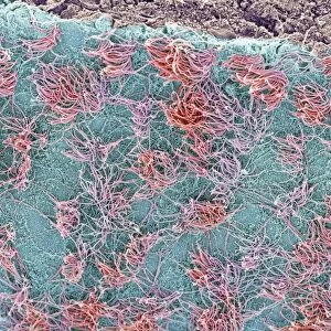
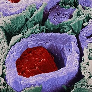
Myelinated nerve, TEM
Myelinated nerve. Coloured transmission electron micrograph (TEM) of myelinated nerve fibres and Schwann cells. Myelin (purple) is an insulating fatty layer that surrounds nerve fibres (axons, green), increasing the speed at which nerve impulses travel. It is formed when Schwann cells wrap around the fibre, depositing layers of myelin between each coil. The outermost layer consists of the Schwann cells cytoplasm (blue/green) and is known as the neurolemma or sheath of Schwann
Science Photo Library features Science and Medical images including photos and illustrations
Media ID 6421804
© THOMAS DEERINCK, NCMIR/SCIENCE PHOTO LIBRARY
Axon Cytoplasm Endoneurium Fatty Fibre Fibres Insulated Insulating Insulation Micrograph Myelin Myelinated Nerve Nerve Fibre Nervous Neurolemma Neuron Neurone Neuroscience Phospholipid Schwann Cell Sheath Sheath Of Schwann Sheathed System Transmission Electron Transmission Electron Microscope False Coloured Neurological Neurology
EDITORS COMMENTS
This print from Science Photo Library showcases the intricate beauty of a myelinated nerve, as seen through a transmission electron microscope (TEM). The image reveals an array of vibrant colors that bring to life the complex structures involved in our nervous system. At the heart of this composition are myelinated nerve fibers, represented in striking shades of green. These fibers serve as pathways for nerve impulses to travel throughout our body. Surrounding these axons is a crucial element called myelin, depicted here in regal purple hues. Myelin acts as an insulating layer made up of fatty substances, enhancing the speed at which these electrical signals can propagate. The process by which myelin forms is truly remarkable and is beautifully captured within this print. Schwann cells, illustrated with their distinctive blue-green cytoplasm, wrap themselves around each fiber like protective sheaths. As they do so, they deposit layers upon layers of myelin between each coil, creating a multi-layered structure that ensures efficient signal transmission. The outermost layer visible in this image represents the neurolemma or sheath of Schwann – it serves as another vital component safeguarding these delicate nerves. This photograph not only offers us a glimpse into the microscopic world but also highlights the incredible complexity and precision found within our own bodies. It reminds us how interconnected biology and anatomy are with neurological processes and provides valuable insights into neuroscience research. Science Photo Library has once again delivered an awe-inspiring visual representation that sparks curiosity about our biological systems while showcasing nature's artistic design at its finest.
MADE IN THE USA
Safe Shipping with 30 Day Money Back Guarantee
FREE PERSONALISATION*
We are proud to offer a range of customisation features including Personalised Captions, Color Filters and Picture Zoom Tools
SECURE PAYMENTS
We happily accept a wide range of payment options so you can pay for the things you need in the way that is most convenient for you
* Options may vary by product and licensing agreement. Zoomed Pictures can be adjusted in the Cart.