Marrow stem, light micrograph

![]()
Wall Art and Photo Gifts from Science Photo Library
Marrow stem, light micrograph
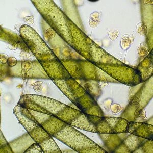
Marrow stem. Light micrograph of a section through the stem of a marrow (Curcurbita sp.), showing the sieve plates in the phloem. A single collateral vascular bundle can be seen. This has an outer and inner phloem (bluish-yellow) with large sieve tubes some of which show sieve plates and companion cells, with an inner xylem (crimson) with large vessels and small woody parenchyma. The cambium (light blue, small cells) is in-between the phloem and xylem. Surrounding the bundle is the ground tissue parenchyma (yellow). Magnification: x37, when printed 10 centimetres wide
Science Photo Library features Science and Medical images including photos and illustrations
Media ID 6338787
© DR KEITH WHEELER/SCIENCE PHOTO LIBRARY
Cambium Cell Biology Cytological Cytology Dicot Dicots Dicotyledon Dicotyledons Ground Tissue Histological Histology Marrow Microscopy Outer Parenchyma Phloem Plates Sieve Plate Sieve Tube Stain Stained Stem Structural Tissue Tubes Vascular Bundle Xylem Cells Collateral Light Micrograph Light Microscope Section Sectioned
MADE IN THE USA
Safe Shipping with 30 Day Money Back Guarantee
FREE PERSONALISATION*
We are proud to offer a range of customisation features including Personalised Captions, Color Filters and Picture Zoom Tools
SECURE PAYMENTS
We happily accept a wide range of payment options so you can pay for the things you need in the way that is most convenient for you
* Options may vary by product and licensing agreement. Zoomed Pictures can be adjusted in the Cart.