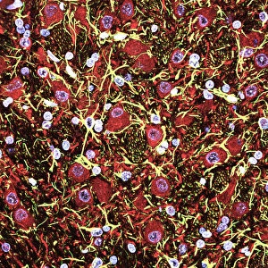
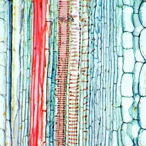
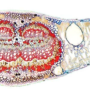
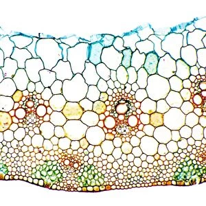
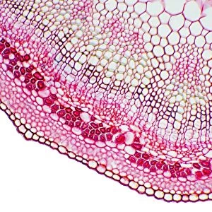
Iris root, light micrograph

![]()
Wall Art and Photo Gifts from Science Photo Library
Iris root, light micrograph
Iris root. Light micrograph of a section through the root of an Iris plant (Iris germanica), showing a vascular cylinder (centre) and parenchyma cells packed with starch grains. The cylinder is comprised of a central cluster of parenchyma cells (red) surrounded by vascular bundles (brown) and the endodermis (white and red ring). The largest vessels seen here are metaxylem, part of the xylem tissue (red) in the vascular bundles. Xylem transports water and mineral nutrients from the roots throughout the plant, while the phloem (green/yellow), the other component of the bundles, transports carbohydrates and plant hormones. Magnification: x100 when printed 10 centimetres wide
Science Photo Library features Science and Medical images including photos and illustrations
Media ID 6302245
© DR KEITH WHEELER/SCIENCE PHOTO LIBRARY
Endodermis Iris Germanica Meta Xylem Monocot Monocotyledon Phloem Plant Anatomy Root Roots Slice Stain Stained Tissue Tissues Transport Transportation Transverse Vascular Vascular Bundle Vessel Xylem Cells Light Micrograph Light Microscope Section Sectioned
MADE IN THE USA
Safe Shipping with 30 Day Money Back Guarantee
FREE PERSONALISATION*
We are proud to offer a range of customisation features including Personalised Captions, Color Filters and Picture Zoom Tools
SECURE PAYMENTS
We happily accept a wide range of payment options so you can pay for the things you need in the way that is most convenient for you
* Options may vary by product and licensing agreement. Zoomed Pictures can be adjusted in the Cart.