Helicotrema cochlear structure, diagram

![]()
Wall Art and Photo Gifts from Science Photo Library
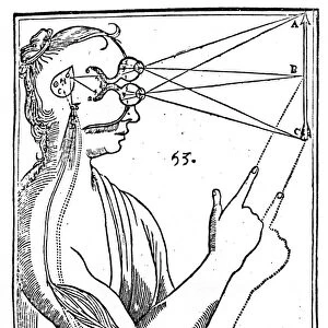
Helicotrema cochlear structure, diagram
Helicotrema cochlear structure. Diagram of a cross-section through the cochlea (an organ found in the inner ear), showing the helicotrema, the place where structures called the scala tympani (orange) and the scala vestibuli (black) meet. Both these structure are perilymph-filled cavities that conduct sound to the scala media (blue), an endolymph-filled cavity that lies between the other two cavities. Within the scala media is the organ of Corti (shown at upper left), which includes inner and outer hair cells (shown at lower left). When moved by sounds waves, these transmit electrical impulses to the cochlear nerve (green)
Science Photo Library features Science and Medical images including photos and illustrations
Media ID 6336556
© FRANCIS LEROY, BIOCOSMOS/SCIENCE PHOTO LIBRARY
Auditory Nerve Aural Cavities Cavity Cochlea Conduction Conductive Cross Section Cut Away Diagram Electrical Impulse Hearing Impulses Inner Ear Internal Nerve Impulse Nerves Neural Organ Of Corti Physiological Physiology Sense Sensory Sound Waves Auditory System Cochlear Nerve Endolymph Nervous System Scala Tympani Section Sectioned
EDITORS COMMENTS
This print showcases the intricate structure of the Helicotrema cochlear, a vital organ found within the inner ear. The diagram provides a detailed cross-section view of the cochlea, highlighting the meeting point known as the helicotrema. Here, two perilymph-filled cavities called scala tympani (orange) and scala vestibuli (black) converge, working in harmony to conduct sound towards the scala media (blue). This central cavity is filled with endolymph and plays a crucial role in auditory perception. The visual representation also reveals another remarkable feature: the organ of Corti located at the upper left corner. Within this sensory apparatus lie both inner and outer hair cells depicted at lower left. These specialized cells play an essential role in converting sound waves into electrical impulses that are transmitted through the cochlear nerve (green), enabling us to perceive sounds. This artwork not only highlights the biological complexity of our hearing system but also emphasizes its normal physiology. By providing a cut-away view into our internal anatomy, it offers valuable insights into how we sense and process sound waves. Delving deep into neural pathways and nervous systems, this image serves as a reminder of just how intricately designed our bodies are to facilitate one of our most fundamental senses - hearing.
MADE IN THE USA
Safe Shipping with 30 Day Money Back Guarantee
FREE PERSONALISATION*
We are proud to offer a range of customisation features including Personalised Captions, Color Filters and Picture Zoom Tools
SECURE PAYMENTS
We happily accept a wide range of payment options so you can pay for the things you need in the way that is most convenient for you
* Options may vary by product and licensing agreement. Zoomed Pictures can be adjusted in the Cart.