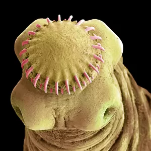
Home > Animals > Fishes > G > Grouper
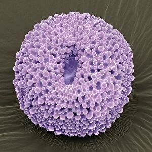
Apoptosis, SEM

![]()
Wall Art and Photo Gifts from Fine Art Storehouse
Apoptosis, SEM
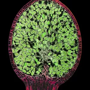
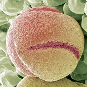
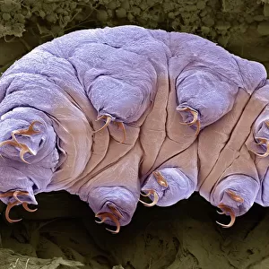
Apoptosis. Coloured scanning electron micrograph (SEM) of a 293T cell in the early stages of programmed cell death, or apoptosis. Apoptosis occurs when a cell becomes old or damaged. The cell becomes spherical as its cytoskeleton, which holds cell shape, is digested, and blebs form on its surface. Eventually the cell breaks into several vesicles, now known as apoptotic bodies, and is phagocytosed (engulfed and digested) by specialist cells. The breakdown into vesicles prevents the leakage of potentially toxic or immunogenic substances from the cell. Magnification: x8000 when printed at 10 centimetres wide.Specimen courtesy of the influenza research group, Professor Wendy Barclay, Imperial College London, UK
Unleash your creativity and transform your space into a visual masterpiece!
STEVE GSCHMEISSNER/SCIENCE PHOTO
Media ID 19527327
© Science Photo Library
Bodies Apoptosing Apoptotic Bleb Blebs
FEATURES IN THESE COLLECTIONS
> Animals
> Fishes
> G
> Grouper
> Fine Art Storehouse
> Science Inspired Art
> SEM (Scanning Electron Microscope)
> Fine Art Storehouse
> Science Inspired Art
EDITORS COMMENTS
This print titled "Apoptosis" offers a mesmerizing glimpse into the intricate world of cellular life and death. Captured using a scanning electron microscope (SEM), the image showcases an early stage of programmed cell death, known as apoptosis. In this snapshot, we witness a 293T cell undergoing transformation as it becomes old or damaged. The cell's cytoskeleton, responsible for maintaining its shape, is being digested, causing the once elongated structure to adopt a spherical form. Notably, blebs start to emerge on the cell's surface – small protrusions that serve as markers of impending demise. As apoptosis progresses further, the cell breaks apart into several vesicles called apoptotic bodies. These tiny remnants are crucial in preventing any potential release of harmful substances from within the dying cell. Specialist cells will eventually engulf and digest these apoptotic bodies through phagocytosis. The vibrant colors added during processing enhance our visual understanding of this microscopic phenomenon while adding an artistic touch to scientific exploration. With a magnification level of x8000 when printed at 10 centimeters wide, every intricate detail comes to life in this stunning piece captured by Steve Gschmeissner from Imperial College London's influenza research group. This print serves as both an educational tool and an aesthetic masterpiece that invites viewers to contemplate the delicate balance between life and death at a cellular level.
MADE IN THE USA
Safe Shipping with 30 Day Money Back Guarantee
FREE PERSONALISATION*
We are proud to offer a range of customisation features including Personalised Captions, Color Filters and Picture Zoom Tools
SECURE PAYMENTS
We happily accept a wide range of payment options so you can pay for the things you need in the way that is most convenient for you
* Options may vary by product and licensing agreement. Zoomed Pictures can be adjusted in the Cart.